
This is the case of a 74-year-old woman treated with 2 overlapping stents in the proximal-to-middle segment of the left anterior descending coronary artery due to severe symptomatic disease. A Sion guidewire (Asahi Intecc, Japan) was recrossed towards the left circumflex artery to finish with postdilatation of the overlapping zone between the proximal left anterior descending coronary artery and left main coronary artery. While the recrossed guidewire was being removed, the distal loop tangled up with the stent at left circumflex artery ostial level and it couldn’t be removed (figure 1A, yellow arrow). A low-profile balloon was advanced over the guidewire that was eventually removed. Still, a small radiopaque distal fragment got trapped in the stent of the left main coronary artery (figure 1B) (Clearstent, Siemens Healthcare, Germany) suspicious of residual material in the coronary sinus not visible through conventional fluoroscopy. The result was assessed on an optical coherence tomography that revealed the presence of guidewire fragments in the left main coronary artery. In-stent postdilatation was repeated with a noncompliant balloon at high pressures (video 1 of the supplementary data). The optical coherence tomography confirmed the crushing of the guidewire fragment on the stent struts (figure 2A, white arrow), and an extremely thin guidewire fragment entering the guide catheter (figure 2B, white arrow, and videos 2-4 of the supplementary data). The procedure was completed, and the patient was discharged uneventfully.
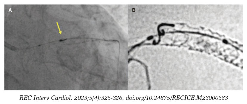
Figure 1.
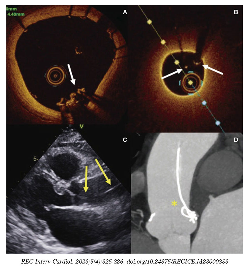
Figure 2.
At 6-month follow-up, an echocardiogram showed a linear hyperechoic image in the ascending aorta (figure 2C, yellow arrows). A computed tomography scan revealed the presence of residual metal guidewire fragments protruding from the left main coronary artery and moving towards the aorta (figure 2D, asterisk). A conservative approach was decided since the patient remained asymptomatic and without inducible ischemia. No clinical events have ever been reported at 2-year follow-up (on dual antiplatelet therapy).
The patient gave her written informed consent for publishing purposes.
FUNDING
None whatsoever.
CONFLICTS OF INTEREST
None reported.
SUPPLEMENTARY DATA
Vídeo 1. Hernández Hernández F. DOI: 10.24875/RECICE.M23000383
Vídeo 2. Hernández Hernández F. DOI: 10.24875/RECICE.M23000383
Vídeo 3. Hernández Hernández F. DOI: 10.24875/RECICE.M23000383
Vídeo 4. Hernández Hernández F. DOI: 10.24875/RECICE.M23000383


