In 2016, 3672 diagnostic intravascular ultrasound (IVUS) studies were performed in Spain. Although the number of cases making use of this technique increased 1.6% compared with 2015, its use has decreased since 2011, when 5124 IVUS studies were performed, in favor of optical coherence tomography (OCT), the use of which has continued to increase.
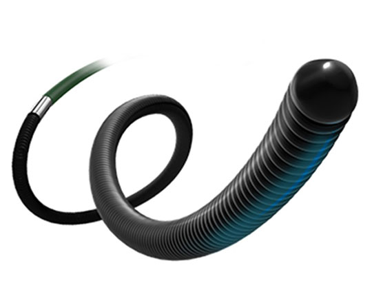
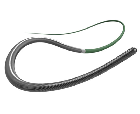
The quality of intracoronary images acquired with IVUS is improving such that it will undoubtedly affect the choice of diagnostic imaging technique. Several systems provide intracoronary ultrasound images with piezoelectric transducers that range from 20 to 60 MHz. In recent years, there has been an improvement in the axial and lateral spatial resolution of intravascular ultrasound. While axial resolution was previously around 40 microns (even with 60 MHz detectors), with the new OptiCross HD system (Boston Scientific), IVUS resolution approaches that of OCT, which can reach 22 microns. The lateral resolution has also improved, from 80-200 to 50-140 microns, again, approaching that of OCT. This advance is due to a change in the design of the piezoelectric transducer of the catheter, which provides a more sensitive signal with less energy loss and a higher bandwidth, improving the resolution while maintaining the penetration of the IVUS.
This improved image quality makes diagnosis considerably easier and means other diagnostic techniques such as OCT, which require contrast to obtain images, are not required. This is highly relevant in certain patients, such as those with compromised renal function.
It remains to be seen how these changes will affect the rate of use of certain diagnostic imaging techniques over others.
Keywords: ultrasound, IVUS, intravascular ultrasound, HD, OCT, optical coherence tomography